How Pain Specialists Evaluate Symptoms: Exams, Imaging, and Nerve Tests
Tests used to diagnose pain include a medical history, physical and neurological exams, imaging, and nerve studies. Premier Injury Specialists matches each test to the pain pattern, symptoms, and suspected cause.
Request an Estimate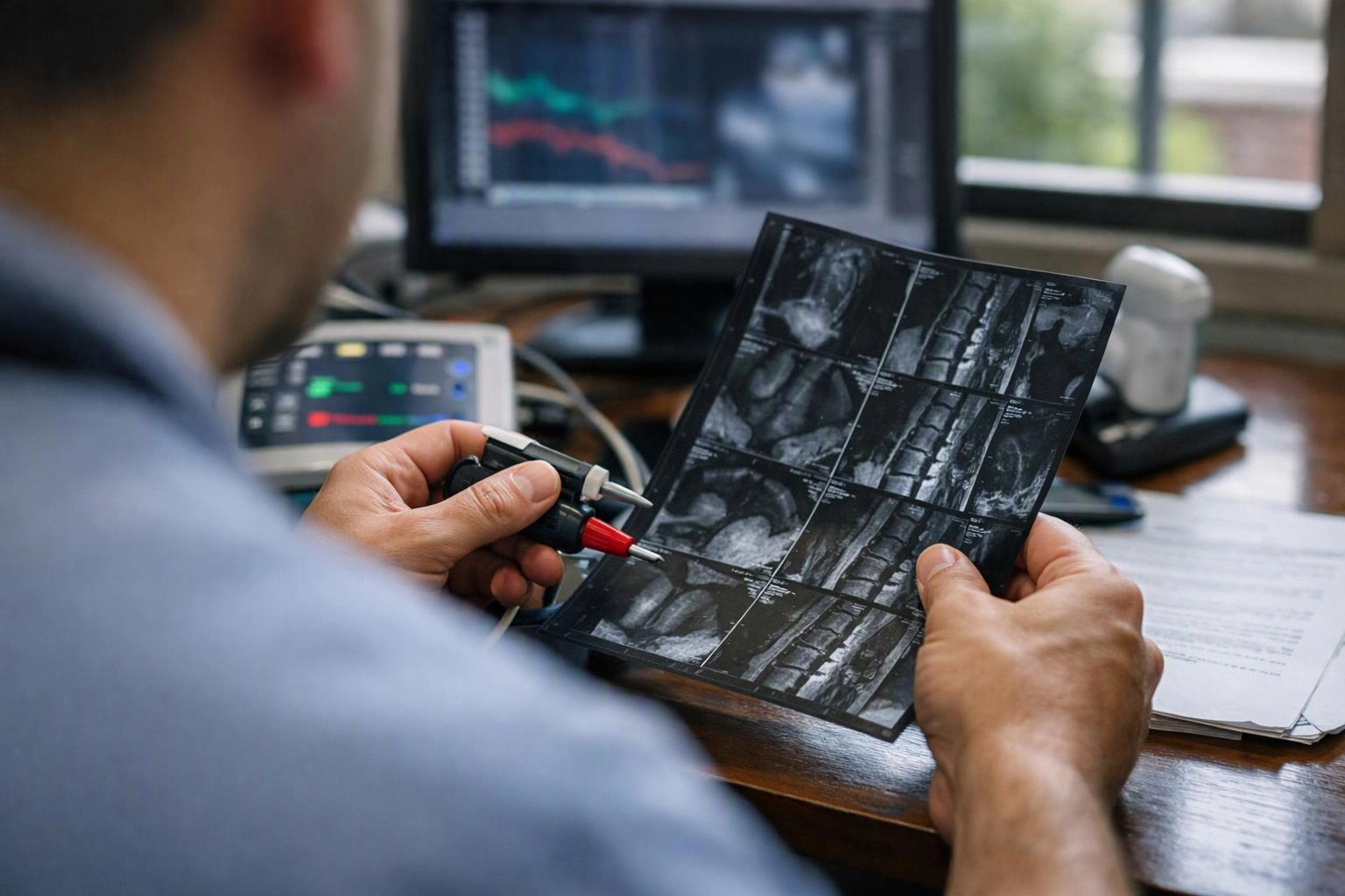
How Does a Pain Management Physician Evaluate Pain?
Pain specialist evaluation connects the symptom pattern with medical history and exam findings. Premier Injury Specialists asks when pain started, where it travels, and what makes it better or worse.
Our team reviews prior injuries, health conditions, medicines, and previous treatment. We also compare the symptom location with signs such as radiating pain, numbness, tingling, or weakness.
A physical examination checks movement and tender areas. A neurological examination checks whether the symptoms may involve nerve irritation or nerve compression.
For patients seeking an auto accident injury evaluation, this process helps connect the injury history with current pain findings.
What Does the Physical and Neurological Exam Check?
A physical and neurological exam checks movement, strength, reflexes, sensation, and signs of nerve irritation. These findings help us decide which diagnostic testing can answer the main clinical question.
We check range of motion in the painful area and compare it with nearby movement. We assess muscle strength and look for weakness that may follow a nerve path.
Reflexes and sensation can show how the nervous system is working. We may also observe gait when back, hip, or leg symptoms affect walking.
When Are Imaging Tests Used to Diagnose Pain?
Imaging tests are used when the exam suggests a structural cause that pictures can help confirm or rule out. Medical imaging can show changes in the spine, bones, discs, and soft tissue.
An X-ray can show bone alignment, joint changes, or other visible bone findings. Magnetic resonance imaging can show discs, nerves, and other soft tissue in greater detail.
Computed tomography creates detailed views of bones and selected soft tissues. We choose a scan based on the symptom pattern, exam findings, and suspected cause.
A scan may show a herniated disc or another spine change. We compare that finding with the symptoms before calling it the pain source.
What Do EMG and Nerve Conduction Studies Show?
Electromyography and a nerve conduction study show how nerves carry signals and how muscles respond to those signals. These tests can help identify nerve damage, nerve compression, or muscle activity changes.
An EMG records electrical activity in selected muscles. The results can show whether a muscle response suggests a problem along a related nerve pathway.
A nerve conduction study measures how signals travel through nerves. Together, the studies can support or question a suspected diagnosis such as radiculopathy.
Why Might a Nerve Test Come Before Imaging?
A nerve test may come before imaging when symptoms point more strongly to nerve dysfunction than to a structural problem. The symptom pattern and neurological findings guide that choice.
Radiating pain, numbness, tingling, or weakness may suggest nerve compression. An EMG or nerve conduction study can help locate the affected pathway before we order a scan.
Diagnostic testing works best when each test answers a clear question. We use the exam and symptom history to choose the most useful starting point.
When Are Both Imaging and Nerve Tests Needed?
Imaging and nerve tests work together when the team needs to connect a visible spine change with its effect on nerve function. Each test answers a different part of the treatment planning question.
These situations can involve magnetic resonance imaging, electromyography, or a nerve conduction study. The combination may help clarify cervical radiculopathy, lumbar radiculopathy, or symptoms linked to a herniated disc.
How Do These Tests Work Together?
Medical history, physical examination, imaging, and nerve studies form a connected clinical evaluation. Our team uses the findings together for treatment planning.
This approach keeps diagnostic testing focused. It also helps us avoid treating every scan abnormality as the source of pain.
What Happens After Pain Testing?
Test results help the pain management team connect symptoms with a working diagnosis and choose the next treatment step. We review the findings with the medical history and examination.
The results may support cervical radiculopathy treatment, lumbar radiculopathy treatment, herniated disc treatment, or sciatica treatment. They may also show that another cause needs attention.
Our team explains what the findings mean and how they affect treatment options. The plan may include continued care, targeted procedures, rehabilitation, or additional evaluation.
For more detail about procedure choices, see Spine Pain Procedures Explained: Epidurals, Facet Injections, and Nerve Blocks. Imaging guidance is also covered in How Imaging Guides Pain Procedures: Fluoroscopy and Ultrasound.
What Should You Bring to a Pain Evaluation?
Bringing complete health information helps the pain evaluation stay focused. These records give our team a clearer view of your symptoms and previous care.
Bring questions about any test already ordered. Our team can explain how each result may affect the next decision.
What Questions Should You Ask About Pain Tests?
Ask what each diagnostic test is meant to show and how the result may guide care. Clear answers can help you understand the purpose of the evaluation.
Ask whether the test checks anatomy, nerve signals, muscle response, or another concern. You can also ask how the findings will affect the working diagnosis and treatment decision.
Ask when and how the test results will be reviewed. Our team can explain the follow-up care connected to those results.
Questions About Pain Exams and Nerve Tests
Can an MRI show the cause of nerve pain?
An MRI can show structural changes that may affect a nerve, such as a herniated disc. Premier Injury Specialists compares the MRI finding with symptoms and exam results before linking it to nerve pain.
What is the difference between an EMG and a nerve conduction study?
An EMG records electrical activity in muscles, while a nerve conduction study evaluates signals traveling through nerves. The studies are often reviewed together to assess nerve function and muscle response.
Do all pain conditions require imaging?
No, all pain conditions don't require imaging. The medical history and examination help determine whether imaging can answer an important clinical question.
What does a normal nerve test mean for ongoing pain?
A normal nerve test means the study didn't show certain nerve or muscle abnormalities at that time. Ongoing pain can still have other causes, so we compare the result with the full examination and symptom history.
Pain evaluation connects symptoms, examination findings, imaging, and nerve studies. Premier Injury Specialists uses that full picture to guide the next care decision.
Related Resources
First Pain Management Appointment: What to Expect and How to Prepare
Compared ToPain Management Cost Factors: What Affects Your Out-of-Pocket Cost
Compared ToInsurance and Pain Management: Verify Coverage Before You Book
Compared ToSelf-Pay Pain Management: Questions to Ask Before Scheduling